Drug Watch
From: 2026-06-29
Nature Medicine - AI Section⭐Promising2 min read
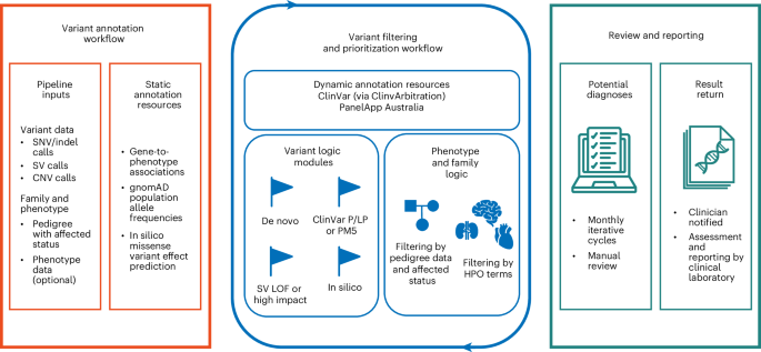
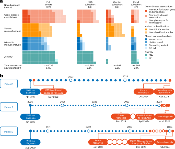
New Free Software Automatically Re-Analyzes DNA to Solve Rare Diseases
Key Takeaway:
An open-source AI tool called Talos automates rare disease genetic data reanalysis, offering a low-cost, scalable way to find new diagnoses as medical knowledge updates.
When patients with mysterious, rare illnesses get genetic tests, the results often do not provide immediate answers. However, as medical science advances, scientists discover new genetic clues every day. This means that re-checking a patient's old DNA data years later can often reveal the correct diagnosis. Because manual re-checking is too slow and expensive for doctors to do regularly, researchers created a new, free computer tool called Talos. Talos automates this entire process, allowing clinics to quickly and cheaply re-analyze patient DNA data on a large scale. This breakthrough could help thousands of families finally find the answers and treatments they have been waiting for.
What this means for you
Scientists created a free tool called Talos that automatically re-checks old genetic tests to find new clues for rare diseases. It is still in development, so patients should not change their care plans.
Citation:
Nature Medicine - AI Section, 2026. DOI: s41591-026-04516-1 Read article →